
A dental centre
at the cutting-edge of technology
At Centre dentaire Jean-François Lévesque, we combine expertise and cutting-edge technology to offer personalized care and lasting results. From sleep apnea to endodontics under a microscope, 3D imaging, and 3D printing of prostheses and dental restorations, we do everything we can to optimize your oral health and comfort.
Sleep apnea
Our team offers specialized dental solutions to improve your breathing at night and reduce the symptoms of sleep apnea. With a custom-made mandibular advancement device, we help keep your airways open, reduce snoring, and promote deeper, more restful sleep.


Digital X-rays
We use Axeos technology to provide high precision 2D and 3D dental images. This imaging allows for more reliable diagnosis and more accurate treatment planning, while reducing radiation exposure. Images are available instantly, facilitating decision-making and improving the quality of our procedures.
Endodontics with an operating microscope
The operating microscope in endodontics allows us to perform root canal treatments with exceptional precision. This tool magnifies the work area up to 25 times, clearly showing the internal structures of the tooth that are difficult to see with the naked eye. This facilitates the removal of infected pulp, cleaning, and sealing of the canals, while preserving the natural tooth. We use this device to provide precise, safe, comfortable, and high-quality endodontic care.


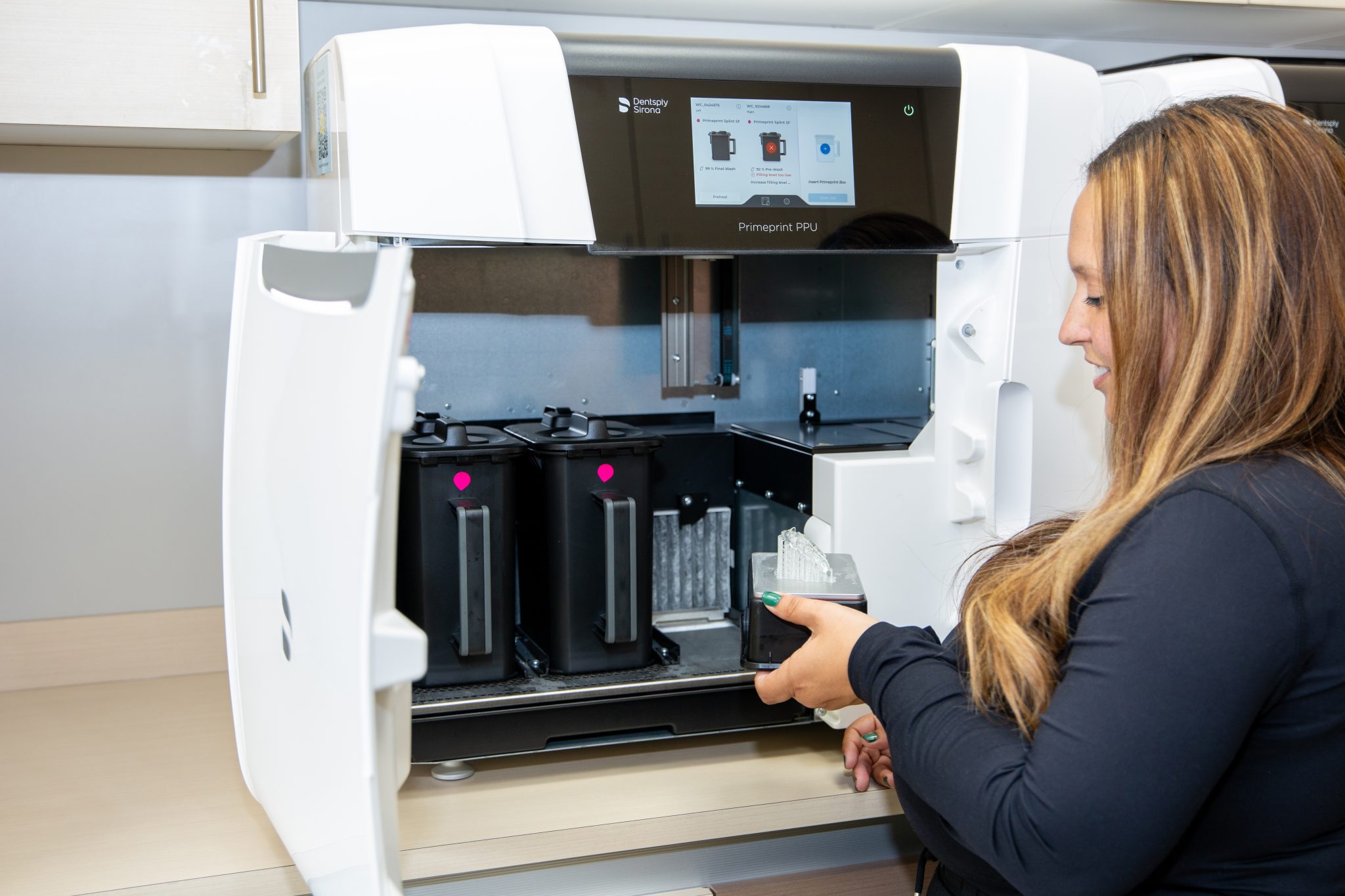
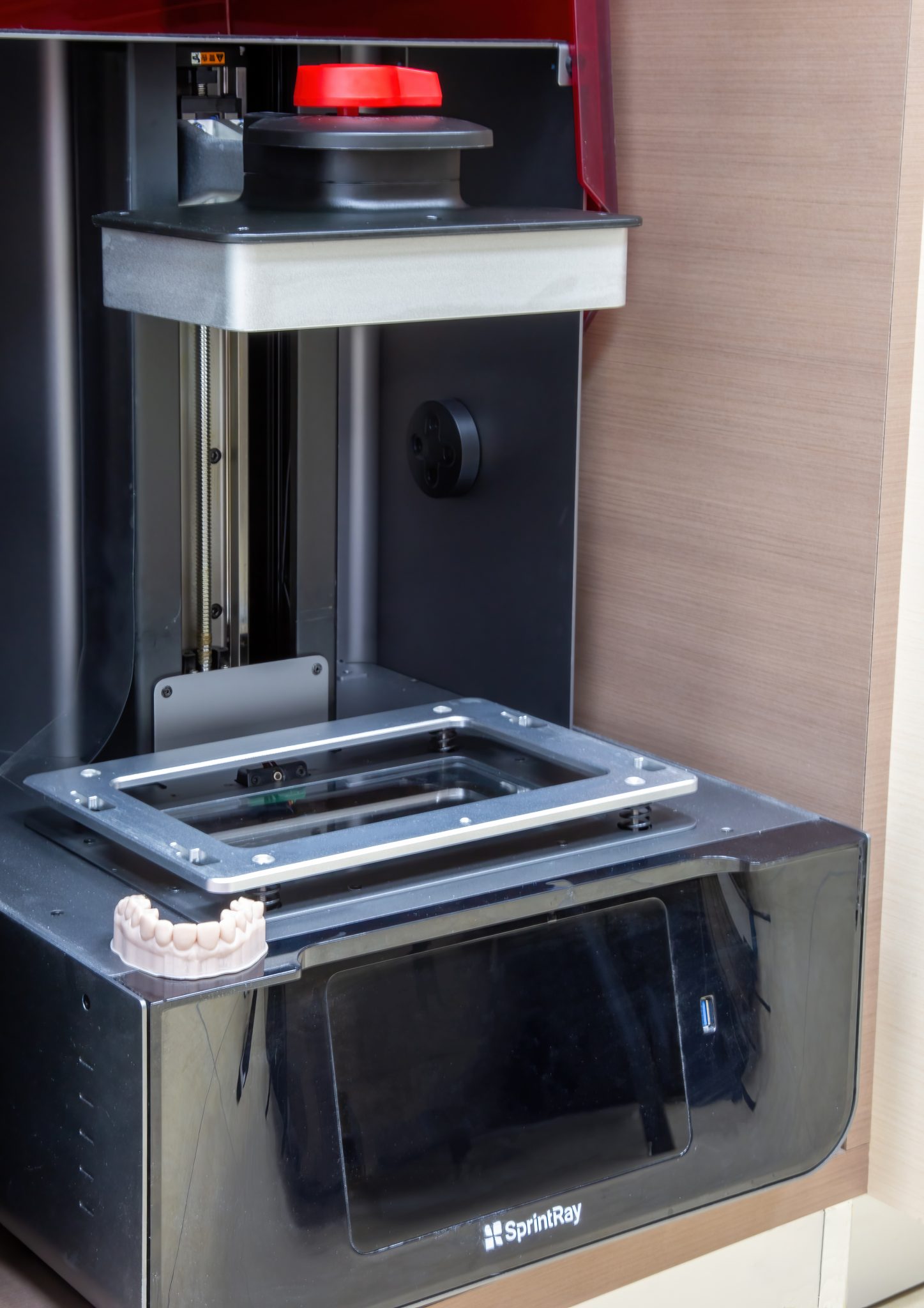
3D printing
We have our own dental laboratory with 3D printing capabilities. We produce highly customized dental prostheses and restorations such as crowns, bridges, veneers, implant pillars, occlusal plates, surgical implant guides, and orthodontic appliances directly on site.
3D printing technology offers greater precision in the design of dental implants and other prostheses. By speeding up the manufacture of prostheses, it can reduce the cost of your dental treatments by eliminating the need for expensive moulds.
T-Scan
The T-Scan is a state-of-the-art tool that accurately records dental occlusion using an electronic sensor, allowing teeth to be adjusted for optimal comfort. T-Scan data helps dentists diagnose and treat a variety of dental conditions related to how your teeth close together (occlusion).
This technology improves the detection of problems invisible to the naked eye and treatment planning. It also makes it easier for patients to understand their occlusion and helps us perform accurate follow-ups. It is used during all our appointments for crowns, bridges, and implants. It complements the coloured papers that were used in the past.


Intraoral camera and LUM
The intraoral camera, a small device about the size of a pen, captures high-resolution images of your teeth and gums, allowing us to identify cracks, cavities, or defective fillings.
These images are displayed on a monitor so that the dentist can examine them and make a diagnosis. This also allows the dentist to show them to you so that you can see the condition for yourself. The camera is connected to a light source, usually a LED, which provides illumination and allows for clear images. The images are also stored on a computer for later viewing and analysis.